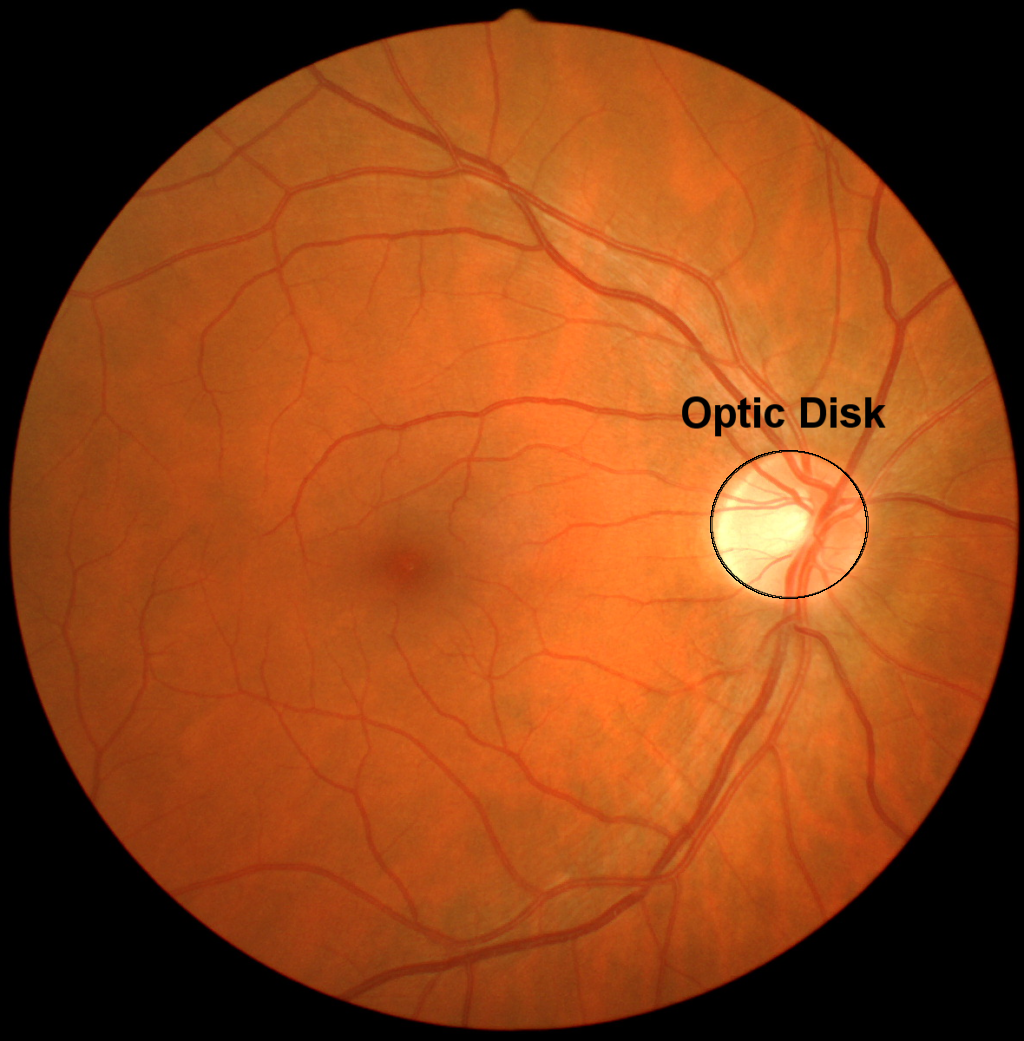
In the photograph below, you can see the retina with the optic disk outlined. The optic disk is the part of the retina where the optic nerve leaves the eye and heads to the brain. In the optic disk, there are no receptor cells. Axons of the retinal ganglion cells gather at the optic disk and form the optic nerve, which carries the neural signal into the brain. Because there are no receptors at the site of the optic disk, this location is also called the blind spot. Because the blind spot on the left eye does not correspond to the same region in space as the blind spot on the right eye, humans really do not have a blind spot in their visual field. Even when viewing the world with only one eye, we are not aware of our blind spot. Higher areas of visual processing “fill in” the blind spot (Ramachandran, 1992).

In this demonstration, you can find your own optic disk and experience this filling in.
To see the illustration in full screen, which is recommended, press the Full Screen button, which appears at the top of the page.
Below is a list of the ways that you can alter the illustration. The settings include the following:
Position: change the position of the x; move it until you cannot see it.
You will need to keep one eye closed.
Size: the size of the x. Can you make
it too large to hide in your blind spot?
Change Eye: which eye you will be using to find a blind spot.
Drawing area: you can also click or touch the
drawing area to move the x.
Pressing this button restores the settings to their default values.